A 3 ml syringe was loaded with lignocaine 2% 1.5ml, Marcaine 0.5%ml and 0.25ml of Garamycin(10mg). The cannula was fitted to the syringe and kept aside.
- The eye was prepared in the routine manner.
- The lid speculum was applied.
- Amethocaine 1% drop was applied every minute for 5 min into the conjunctival sac.
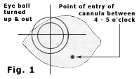
 The patient was instructed to look upwards and outwards. In the infra-nasal quadrant , 5 mm away from the limbus, between four and five o’clock position, the conjunctiva was cauterized over an area of about 2mm in diameter (Fig. 1).
- The cauterized conjunctiva was held with a corneal forceps, and a nick was made in the conjunctiva with a Wescott-syle scissors. This exposed the pearly white Tenon’s facia.
- The Tenon’s facia was held with a toothed corneal forceps. A nick was made in the Tenon’s facia. The gap in the Tenon’s facia exposed the bare sclera. About 5 mm of the tip of the scissors was introduced into the opening of the Tenon’s space. This facilitated introduction of the cannula into the sub-Tenon’s space.
 The cannula was positioned in such a way that the curvature of the cannula conformed with curvature of the eye ball. The tip of the cannula was placed in the opening (Fig 2). If the eye ball moved, the eye ball was stabilized by holding firmly with a toothed forceps, close to the limbus at either four or five o’clock position.
- The cannula was gently pushed making sure that the tip of the cannula was in close proximity to eye ball as it was pushed behind the eye ball.
 When the cannula was pushed in, there was resistance in some eyes, owing to scleral-Tenon bridging fibres near the equator of the globe. When there was any resistance, a small amount of the anesthetic solution was pushed in. This hydrodisected the resisting tissue. Then the whole curved part of the cannula was pushed into the sub-Tenon’s space. At this point, the remaining 0.5mm straight portion of the cannula and the syringe were radial to the eye ball. The anesthetic solution was gently emptied into the sub-Tenon’s space. In some eye, the initial resistance was high. Once the solution entered, the resistance became less. The solution was pushed slowly and gently till the syringe was empty. The cannula was pulled out gently in the curved path it entered.
- The surgery was performed after 10 minutes.
